Xero Image Technologies is an authorised agent for ADANl, who offer a extensive range of medical products. Below are some examples of scans by the ADANI Range.
In comparison with general-purpose digital radiography systems based on flat panel X-ray imaging receivers, the UNIVERSAL digital radiography system provides a considerable number of clear advantages for performing speedy diagnostics of patients.
UNIVERSAL is designed to obtainig standard size high-quality x-ray images of the patient’s bone structures and soft tissues in the standing, sitting and lying positions.
Intended Use
The UNIVERSAL is designed for obtaining standard size high–quality x–ray images of the patient's bone structures and soft tissues in the standing, sitting and lying positions.
The main features and benefits
Low dose. The overriding advantage of digital scanning technology is that digital images offer consistently excellent image quality at a lower dose.
Cost reduction expenses. No film costs as well as improving workflow and increasing throughput can have a dramatic impact on expenses. Reliable and sturdy design reduces maintenance expenditure.
Compactness. The UNIVERSAL offers a compact system with excellent features. You can even create a standard radiography room in smaller than average rooms in particular where the ceiling height is lower than average and othere systems will not fit.
Speed and safety. The UNUSCANsystem gives both operators and patients fast and effective solution with a maximum level of performance.
Sophisticated image processing technology. ADANI have developed a solution that allows all diagnostically relevant information to be displayed in one single image, giving you more confidence in diagnosing. Manual procedures, like applying wedge filters, multiple processing to display all requested areas of the image or adjusting contrast and brightness, become obsolete.
Digital integration saves time. The automated digital workflow allows for the timesaving handling of patient and administrative data via RIS and PACS integration. Also shortened image acquisition times and faster image distribution processes increase departmental efficiency. Shorter waiting times for the patients increase customer satisfaction. This enables operators to spend the saved time with the patients.
The main advantages of slot-scan chest radiography are the low cost, the low radiation exposure and the speed at which chest imaging can be acquired and interpreted. The design of the PULMOSCAN system meets the specific screening requirements such as high patient throughput and , fast viewing in a compact and sturdy design.
The PULMOSCAN is designed and used for the examination (chest screening) for the early diagnostics of tuberculosis, oncological and other diseases withinthe chest organs.
Intended Use
The PULMOSCAN is intended to produce high quality digital radiographic chest imaging for the purpose of screening and diagnostic chest radiography.
PULMOSCAN
Designed specifically to to perform early diagnostics of the diseases of the thorax organs including tuberculosis, cancer and others
optimal solution for equipping of radiology departments of regional outpatient clinics, hospitals and diagnostic centers
low operating costs as there is no need in X-ray film, chemical, film processor, etc.
Medical and productivity benefits:
Cost-effective solution. The main advantages of slot-scan chest radiography are the speed at which chest imaging can be acquired and interpreted, the low cost and the low radiation exposure
Reduced radiation dosage. Digital radiography achieved comparable image quality with conventional film radiography with a 50-70 percent radiation dosage reduction
Increase image quality.
Increase image throughput. Thereby reducing the cost of operations and improving radiologist productivity
Ease of handling.
Facilitation of the development of a teleradiology network.
The Mobile digital X-ray chest screening is in fact the best and possibly the only method available to reach the worst affected groups. It helps raise awareness of TB among high risk groups and those who work with them.
Adani have developed The PULMOEXPRESS mobile unit is used to actively identify cases of TB at an early stage of the disease progression, before someone becomes infectious and can pass the disease to other people.
PULMOEXPRESS is designed for chest screening various population groups for early diagnostics of tuberculosis in rural areas, or in military units, refugee camps or in penitentiaries.
We recognise that local markets may require a different specification and are experienced at working with national and international companies to develop a Mobile Chest Radiography Unit that meets the particular needs of different markets.
Adani can work with you to provide a suitable design that meets th needs of your particular market requirements.
This solution provides a self-contained mobile PULMOSCAN chest radiography unit inside the van.
Intended Use
PULMOEXPRESS
Intended to produce high quality digital radiographic chest imaging for the purpose of screening chest radiography,
Intended to perform digital chest imaging on the target groups identified as being at high risk of TB including: homeless people, prisoners, drug users and street-drinkers, refugees.
The PULMOEXPRESS capable of safely screening 300-350 people a day. The screening process is quick taking 2 — 3 minutes per person and the results are given immediately.
Design Features
The compact cost-effective solution of the van
Designed to look internally like a standard medical office
Equipped with the most advanced PULMOSCAN chest digital radiography system
Single-phase power supply
High resistance to transport vibration
High resistance to temperature variations when not operating
Key Benefits of Mobile Chest Radiography Screening
High throughput
Low dose
Better patient compliance
Preventive exams close to home
Early detection substantially improves survival rates
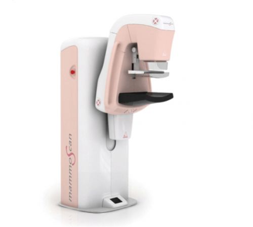
The MAMMOSCAN FFDM system offers the industry's highest image resolution (48 µm) with dramatically reduced radiation dose and the virtual elimination of motion artefacts.
The MAMMOSCAN is intended to produce standard size high-quality x-ray images of the human breast for the purpose of diagnostic and screening mammography.
The MAMMOSCAN utilizes a slot-scanning imaging technique, with a thin X-ray beam scanning across the breast while a multi-linear array detector is moving under the breast.
This FFDM system with leading-edge design and high performance with low dose radiation exposure.
Intended Use
The MAMMOSCAN is a full-field digital mammography X-ray system, designed to perform digital X-ray breast imaging for diagnostic and screening purposes (i.e., for early breast cancer detection).
MAMMOSCAN is designed to be used in clinical practice to the same purpose, as a traditional analog (film-type) mammographic apparatus.
Design Features
Digital scanning receiver of X-ray image with 54 microns pixel size
Anti-scatter grid free design, allowing for patient dose reduction with no loss of image quality
High loading factor of X-ray tube, providing long-term stable operation together with high throughput and efficient patient examinations
Compact and ergonomic system design
Motorized C-arm height and rotation adjustment
Touch-screen interfaces at the operator workstation
Two types of replaceable compression paddles of standard dimensions, used depending on the breast size
Digital indication of breast compression force, compressed thickness and projection angle
Advantages
In comparison with digital mammographic apparatus based on standard flat panels, image plates and other types of digital imaging receivers, the digital mammography scanning system ADANI MAMMOSCAN provides a considerable number of clear advantages for performing speedy lower dosage diagnostics of patients.
High spatial resolution MAMMOSCAN generates an image size of 4096 pixels x 5560 pixels with a 22 cm x 28-cm field-of-view, a pixel size of 54 microns with a limiting resolution of 10 line pairs/mm in standard-resolution mode
High contrast resolution High contrast resolution together with ability to manipulate images makes detection of breast cancer more accurate
Low dose The total scan time for the 28 cm image width is less than 6 seconds, with an effective exposure time of 0.4 seconds. The slot-scan technology results in minimized scatter radiation. The faster examination may induce more women to comply with screening at recommended intervals, thus making mammography facilities more efficient.
High throughput Powerful X-ray tube assembly unit with a dedicated heat exchanger employed provides the higher patient throughput within a busy clinical practice
Enhanced image processing The shielded area incorporates the operator´s workstation. The MAMMOSCAN is designed with open architecture for greater compatibility with existing network environments
The MAMMOEXPRESS mobile digital mammography unit has the potential to reduce breast cancer morbidity and mortality by improving access to screening for those hard to reach groups of rural women, busy women, and medically-underserved populations. It is also an opportunity to bring subspecialty breast imaging expertise to more of the population.
We recognise that local markets may require a different specification and are experienced at working with national and international companies to develop a Mobile Mammography Unit that meets the particular needs of different markets.
The self-contained mobile mammography clinic can provide the complete mobile clinic solution, featuring the latest in state-of-the-art digital mammography technology and service. Mobile digital mammography has the potential to reduce breast cancer morbidity and mortality by improving access to screening for rural women, busy women, and medically-underserved populations. It is also an opportunity to bring subspecialty breast imaging expertise to more of the population.
Intended Use
MAMMOEXPRESS is a mobile mammography clinic, designed to perform digital X-ray breast imaging for the purpose mobile mammography screening (i.e., for early breast cancer detection).
Design Features
Compact cost–effective solution of the van
Equipped most advanced MAMMOSCAN FFDM system
Single-phase power supply
High resistance to transport vibration
High resistance to temperature variations when not operating
TRAUMASCAN is intended for obtaining whole body (overview) digital x-ray images of injured patients both in AP and LP projections for the purpose of x-ray diagnosis and quick rendering of qualified medical aid in cases of poly-trauma injuries.
TRAUMASCAN
Is a whole-body digital radiography system, designed to produce the digital X-ray images with a flexible format (from whole- to partial-body areas) of injured or surgical patients where rapid diagnosis of polytrauma is required
It provides critical life-saving information to the medical staff by enabling them to have a complete picture , from head to toe, of a patients injuries, faster and with less interference, due to the patient stabilization features, than was previously possible
Is intended to be used in emergency departments, trauma centers, trauma resuscitation units, radiology departments of clinics, military/ emergency mobile hospitals
DIGITAL X-RAY IMAGING TECHNIQUE
The TRAUMASCAN system is based on enhanced linear slot-scanning technology which produces digital radiographic images in a flexible format and selectable spatial resolution in seconds. The quick X-ray diagnostics are based on performing the following kinds of examinations.
The whole-body X-ray images of the patient to is obtained by stitching together the two large-format images with the 100 cm x 49 cm size are obtained by the two subsequent scans of the lower and upper portions of the patient’s body each taking approxuimately 8 seconds. The time period between the first and second scan should not exceed 20-30 seconds. Resulting in a whole body X-ray in under 1 minute.
A large-format X-ray images of any selected area of the patient’s body with the dimensions of up to 100 cm х 49 cm is obtained as a result of the longitudinal scanning of the patient with the X-ray beam taking 5 — 8 seconds.
High-resolution partial-body X-ray images are obtaining by using standard-format (35 cm x 43 cm) flat panel X-ray receiver or CR imaging plates.


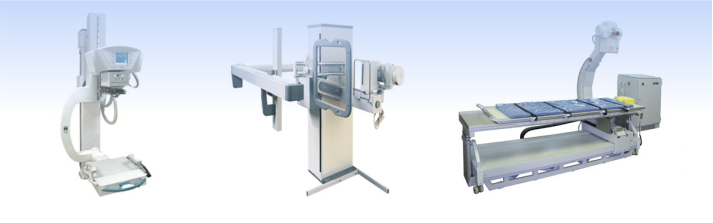
 Multipurpose Digital Radiography Systems